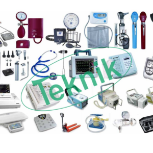
Scientific instrumentation industry has been playing a key role as a basis for technological development to sustain the growth of the world, with the passage of time there is an ever-increasing need for us at Microteknik to meet the more diversified and sophisticated demand from all walks of our industrial society. Microteknik with enormous and diverse experience, are one of the leading manufacturer, exporter, dealer and supplier of scientific laboratory equipment and instruments Since 1973. With an aim to meet the overwhelming demand of the scientific community, we provide an entire range of instruments right from basic lab equipment to most sophisticated instruments for research labs.

ABOUT MICROTEKNIK
Our Skills
Company Profile
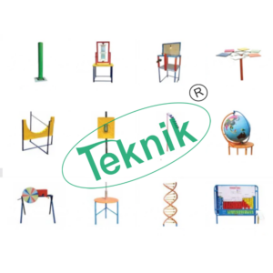
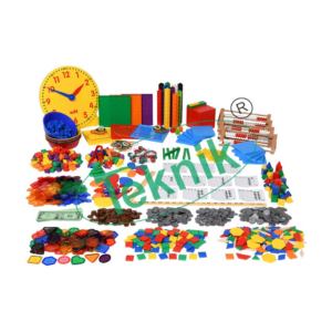
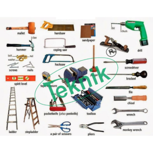
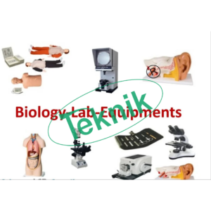
We MICROTEKNIK, an India based manufacturer of all types of world class quality of Scientific Equipments, Laboratory Instruments, Educational/Technical teaching equipments (for school, colleges, universities and vocational training institutes), Hospital equipments, Rehabilitation equipments, Agricultural equipments, Mechanical, Mechatronics, Electical and Electronics equipments, Blood bank Equipments, Automotive-Automobile Lab Equipments, Glassware, Plasticware and Anatomy Models.
Microteknik also produce tailored made equipments as per client specifications / custom built products.
We carry with us experience of over 5 decades, 3rd Generation in the field. Microteknik has carved out their niche in customized products, developments, handling of complete supply, installation, commissioning, training and handling over projects in time with value added services of rich experience.
Concept is not only supply but to ensure customer delight with unsolicited services by means of assisting client during installation and commissioning. Periodic after sales service visits.
Microteknik, a qualified and experience team with QC system and quest for continual improvement.
We stand committed for right quality product with right price, timely delivery with support on installation, commissioning and training at site. Thus making it Single Point Shop, with this today Microteknik is one of the leading manufacturers and exporter in Northern part of India and earned the name as “preferred partner” to the clients.
Microteknik housed in spacious area, equipped with all latest high precision automats and machineries, to cater any bulk requirement supported by highly skilled experince work force and engineers.
Our website www.microteknik.com, www.microteknik.net and www.engineeringcollegeinstruments.com will give you complete product range.
We value your relationship with us and we assure you of our best services at all the times.
Best Regards,
Mr. Vikas Jain
Director (International Sales)
Mobile No.: +91-8930344845
E-mail: info@microteknik.com, microteknik@microteknik.net
Regd Office: 2759, Timber Market, Ambala Cantt- 133001 (India)
Factory: 1, Teknik Tower, Vikaspuri, Main Road, Industrial Area, Ambala Cantt-133006, Haryana (India)
73, Teknik Eatate, Vikaspuri, Main Road, Industrial Area, Ambala cantt-13306, Haryana (India)
Why Microteknik
MICROTEKNIK have successfully earned and retained a strong position as a leading Manufacturer, Exporter, Dealer and Supplier, our precious clients trusted on our high quality and timely shipment. We are following reasons to preferred choice of our clients:
High Quality Standards
Highly Professional Team
Organized Network
Easy accessibility
Affordable prices
Timely Shipping
Products Quality
We are too much conscious about quality, we are manufacturing all types of Scientific Laboratory products in full of dedication to the defined all international and national industry norms and standards. We inspect all material we use from raw material to finishing mode. We are following Advanced TQM Policy in all business operations. Our Quality Analyst Department checked all products with advanced quality testing-equipped with ultra-modernized tools. Our Parameters on which we test our products as following:
Flawlessness
Durability
Dimensional Accuracy
Customized Design
Performance
User-friendly
Our Team
Microteknik have deployed a highly dedicated team of professionals which enable us to manufacture and supplies wide range of engineering and scientific laboratory equipment in an efficient manner. Our all team member is highly experienced and professional of his respective field. We are professionals in:
- Engineers
- Managers
- Technical Experts
- Quality analysts
- Packaging experts
- Sales and Marketing Personnel
- Finance
- R&D personnel
- Semi-skilled laborers
- Warehouse personnel
- Administration personnel
- Technocrats
- Logistic personnel
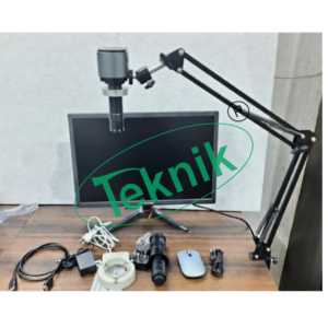
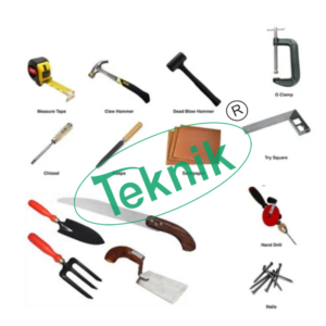
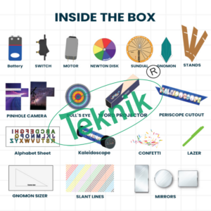
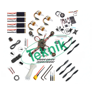
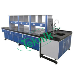
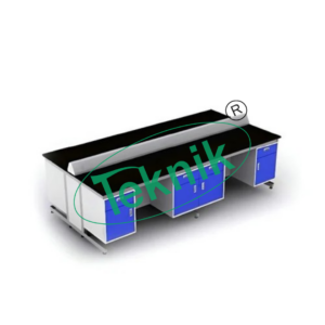
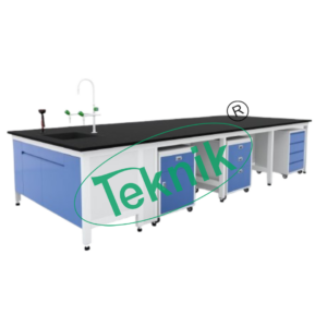
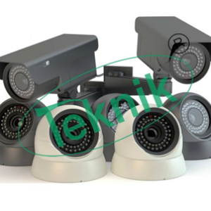
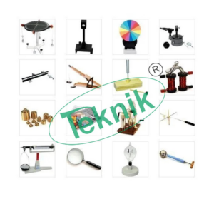
Latest Products